LJI Postdoctoral Associate Dawid Zyla, Ph.D., is a skilled infectious disease researcher. As a member of the Saphire Lab, Dr. Zyla uses a technique called cryo-electron microscopy to capture 3D images of viruses—and their weaknesses.
In cryo-electron microscopy, researchers freeze a very small sample and then use LJI’s cutting-edge cryo-electron microscopes to pelt the samples with electrons. As they strike the samples, the electrons in the sample emit energy, letting researchers see them like never before. The Saphire Lab has used this method to reveal key therapeutic targets on SARS-CoV-2, Ebola virus, and more.
Dr. Zyla is also a talented photographer. He recently captured the key steps in his work to share, in his own words, the technical marvels behind biomedical research.
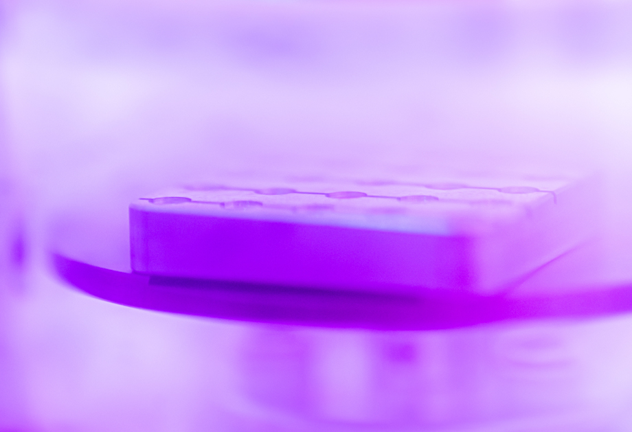
Beginning in plasma
Dr. Zyla: “In cryogenic electron microscopy, everything starts in plasma. To prepare a sample, grids with small pieces of copper covered with a thin amorphous carbon film are cleaned and coated with charged particles. This is called the ‘glow discharge’ step. We do this work in a device called a plasma cleaner, which is connected to a vacuum pump that allows us to have low gas pressure in the chamber. Next, I apply a current between the electrodes that cause glow discharge: a plasma made of charged gas particles. Plasma present in the chamber can be observed as a purple glowing effect (in the picture). Glow discharge is required to remove residual particles present on the grid surface and make them more hydrophilic, which helps us capture a clear image later in the process.”
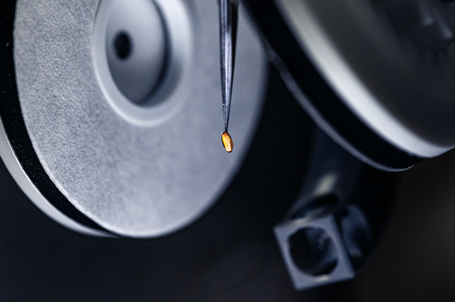
Art of blotting
“Most of the cryo-EM samples are proteins in an aqueous solution, so we need to blot them to remove excess fluid. Manual blotting is a highly imprecise method. Thus various automated devices have been developed to standardize the task. Here is a close-up picture of one of them: a Thermo Vitrobot Mark IV. Vitrobot has a humidity-controlled chamber with two blotting pads and tweezers to hold the grid for the sample application (in the picture). Good blotting conditions are tricky to get, and we usually have to try several times before we get it right. The blotted grid is quickly transferred to a container filled with liquid ethane placed below the humidity chamber and flash frozen. Fast freezing is a crucial step in cryo-electron microscopy.”
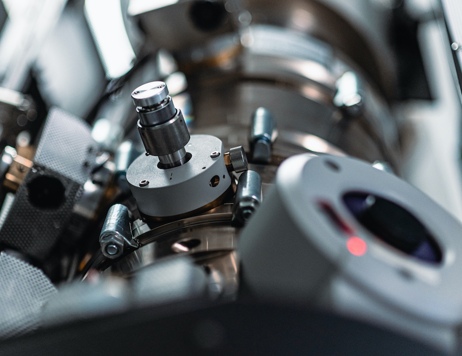
Screening first
“Preparing cryo-electron microscopy grids is knotty and it is relatively easy to make poor quality grids. It gets even harder when we factor in protein samples and their quality. Before we can solve any protein structure, we have to check grid quality using a cryogenic transmission electron microscope. This is the Titan Halo. The data we get from this microscope help us optimize samples and solve low-resolution reconstructions—which guides us toward other protein structures to pursue.”
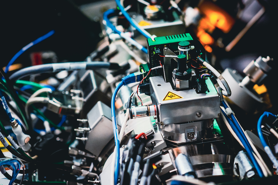
King of them all
“And there it is, the mightiest of them all. Thermo Titan Krios. The workhorse of high-resolution cryo-electron microscopy. This microscope is fully automated yet requires highly trained staff. It provides the highest quality of data possible with a blazingly fast direct electron detector. These technologies allow LJI scientists to solve macromolecule structures that would never be possible to tackle with other methods. The Krios works almost 24/7 and enables us to collect over 6,000 micrographs, images of the sample at very high magnification, per day.”
Related video: Using Viruses to Monitor the Efficacy of the Immune System