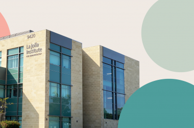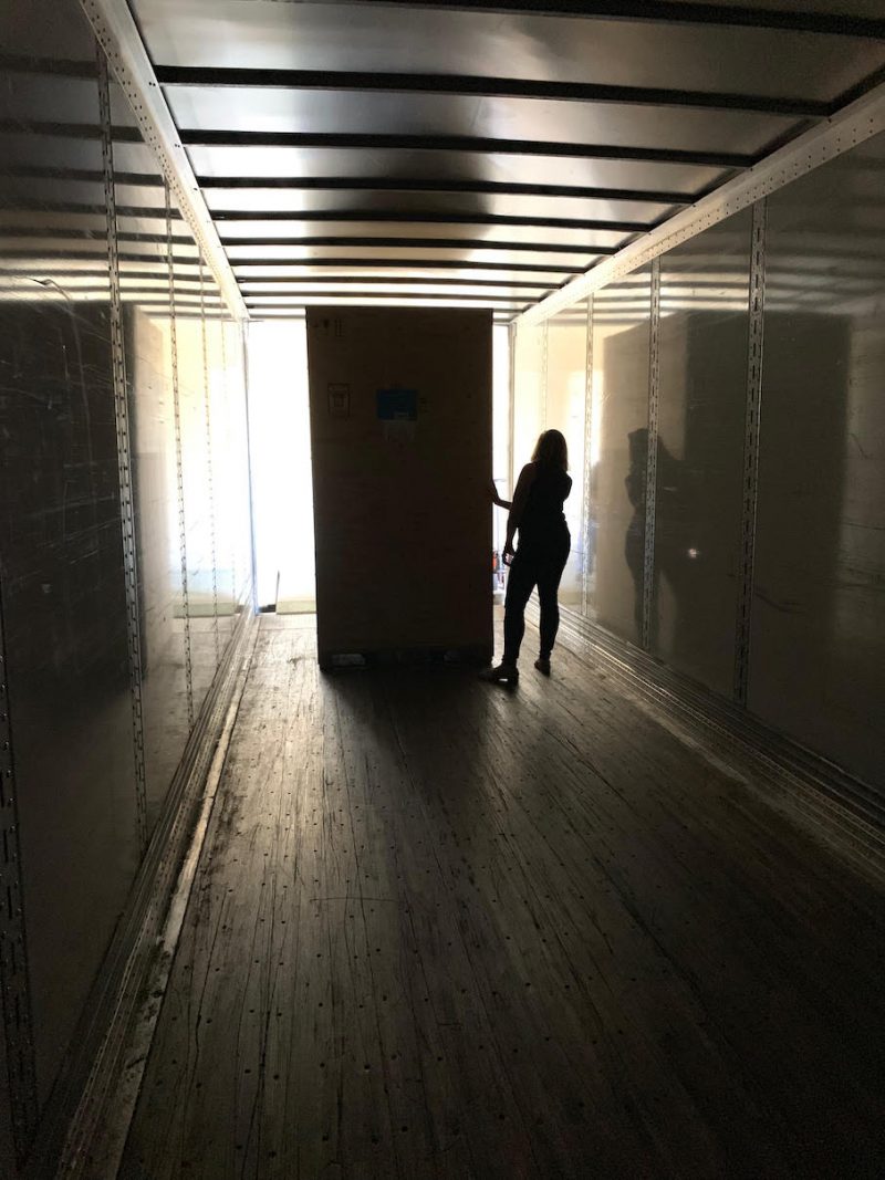
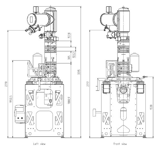
During the last week of August, the trucks finally started to arrive. Carefully nestled inside 22 huge wooden crates was what’s casually referred to as the Krios—shorthand for the world’s most advanced cryo-electron microscope. The Krios is the cornerstone of what soon will become a state-of-the-art molecular imaging facility at La Jolla Institute.
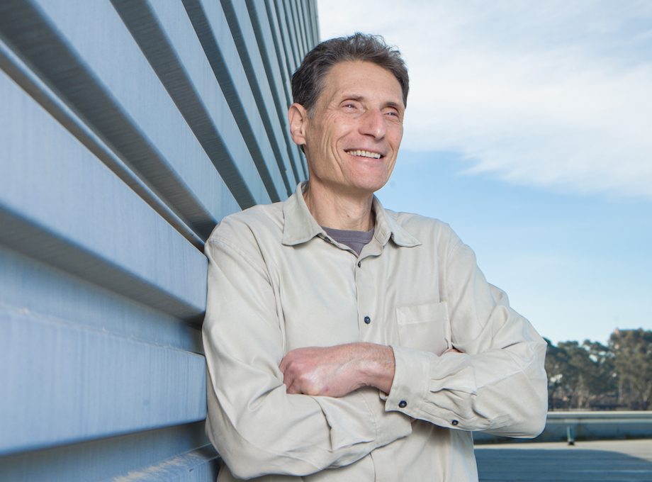
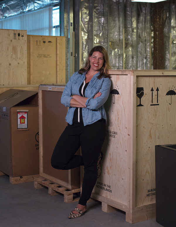
“What’s most remarkable is that it has been barely a year since LJI and I started talking about the idea of building a cryo-EM facility,” said Professor Erica Ollmann Saphire, Ph.D., a widely recognized structural biologists and one of the world’s leading experts in pandemic and emerging viruses, who joined LJI faculty this spring. “What was a mere concept back then has progressed to assembly of the microscope in a newly constructed facility, and electrifying opportunities.”
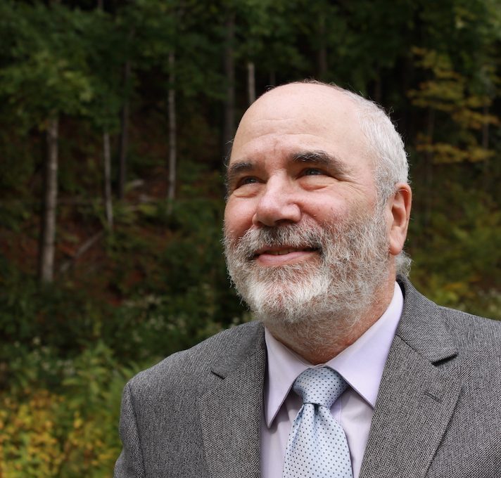
“We are thrilled to welcome Erica to the Institute. She is a truly exceptional scientist who uses molecular insights to bring together scientists and policymakers for scientific advancement and social change,” says Mitchell Kronenberg, Ph.D., LJI President and Chief Scientific Officer. “Having her here will accelerate the Institute’s efforts to solve humanity’s most pressing health challenges, and continue to elevate the remarkable science on the Torrey Pines Mesa.”
Dr. Ollmann Saphire’s decision to move her lab to La Jolla Institute was, in part, driven by the opportunity to build a world-class molecular imaging facility. For decades, biologists have relied on x-ray crystallography— blasting x-rays at crystallized proteins—to determine the structure of biomolecules. But over the last couple of years, cryo-EM has taken the world by storm.
Cryo-EM had long been derided as “blobology” for the indistinct images it delivered, but a perfect storm of improvements in camera technology, image processing, reduced cost, and increased computing power has transformed cryo-EM from low-resolution technique to the most important new way to gain high-resolution insights. Crucially, cryo-EM can also visualize moving parts.
Cryo-EM uses electron beams to image flash-frozen biological molecules and lay bare their threedimensional shapes. Not surprisingly, labs around the world are racing to adopt cryo-EM, because it can provide scientists with a blue print of large, complex proteins’ structures that can’t easily be formed into large crystals.
At the heart of all advances in immunology is the fundamental understanding of the molecular interactions that drive health and disease,” says Dr. Ollmann Saphire. “The extremely detailed images produced by cryo-EM reveal precisely how essential mechanisms of the immune system operate and what goes wrong when one player is out of tune.”
As part of her own work, Dr. Ollmann Saphire studies the three-dimensional structures of viral proteins to understand, at the molecular level, how viruses like Ebola, Marburg, and Lassa are so deadly. In a series of groundbreaking discoveries, her team has identified the molecular structures of the Ebola, Sudan, Bundibugyo, Marburg, LCMV, and Lassa virus surface glycoproteins; how these viruses suppress immune function; and where human antibodies dock to defeat these viruses. This was the roadmap science needed to develop potent therapeutics and vaccines.
She showed that certain viral proteins are surprisingly dynamic and can twist themselves into new shapes at critical stages during a virus’ life cycle to perform different functions. The discovery of what she calls “transformer” proteins expanded the central dogma of molecular biology, which states that a protein’s sequence determines its one-and-only shape and thus its function.
Dr. Ollmann Saphire is also the galvanizing force behind the Viral Hemorrhagic Fever Immunotherapeutic Consortium (VIC). Under her leadership, the global consortium—now headquartered at La Jolla Institute— brings together the expertise of leading structural biologists, virologists, immunologists, clinicians, and public health practitioners to effectively combat disease in humans infected with Ebola and other related filoviruses; arenaviruses such as Lassa virus; as well as a third major global threat, alphaviruses, which infect millions of people across the world.
“The recent resurgence of Lassa, the difficulties in containing Ebola outbreaks, and the re-emergence of alphaviruses in multiple locations, including the United States, makes the development of therapies against these threats an urgent local and global concern,” says Dr. Ollmann Saphire.
Earlier this year, the VIC was awarded up to $35 million by the Centers of Excellence for Translational Research (CETR) program at the National Institute of Allergy and Infectious Disease to continue the tremendously successful international program for another five years.
Dr. Ollmann Saphire’s work has been recognized at the White House with the Presidential Early Career Award in Science and Engineering, with young investigator awards from the International Congress of Antiviral Research, the American Society for Microbiology, and the MRC Centre for Virus Research in the United Kingdom. She has also been recognized with an Investigators in the Pathogenesis of Infectious Disease Award from the Burroughs Welcome Fund, and by the Surhain Sidhu award for the most outstanding contribution to the field of diffraction by a person within five years of their Ph.D.
She has been awarded a Fulbright Global Scholar fellowship from the U.S. Department of State and a Mercator Fellowship from the German Research foundation, Deutsche Forschungsgemeinschaft, to develop international collaborations around human health and molecular imaging through cryo-EM.