Each of our organs is like a special little habitat. Immune cells need to adapt to this habitat in order to defend it from infections, tumors, and other threats.
Some organs, like the liver, are more acidic. Some organs, like the lungs, come into constant contact with pollen, pollution, and pathogens from the outside world. And each organ has a different structure.
Scientists at La Jolla Institute for Immunology (LJI) are investigating which kinds of immune cells take the lead in defending different organs. LJI Assistant Professor Miguel Reina-Campos, Ph.D., has been working with experts in LJI’s Microscopy and Histology Core to capture stunning images of immune cells in mouse tissues for a new research tool called ImmGenMaps.
ImmGenMaps is a great example of how LJI scientists advance medical research. The project is part of the Immunological Genome Project (ImmGen), founded and supported by the National Institutes of Health’s National Institute of Allergy and Infectious Diseases. The initiative unites leading researchers from immunology and computational biology labs around the globe.
“This work is crucial to understanding how long-term immune protection is achieved across tissues with markedly different architectures,” says Dr. Reina-Campos.
Kidneys
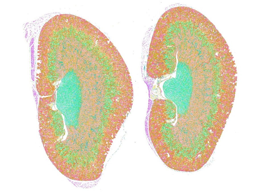
This cross-section of two mouse kidneys features different colors to indicate different cell types. This image is part of an important project to understand how immune cells activate and move through organs over time: ImmGenMaps. This field of research, called spatial transcriptomics, gives scientists an incredibly detailed look at how the body fights disease.
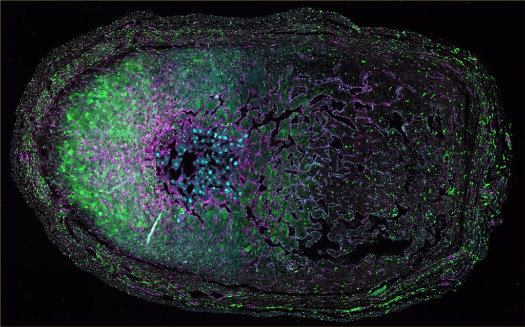
Lung
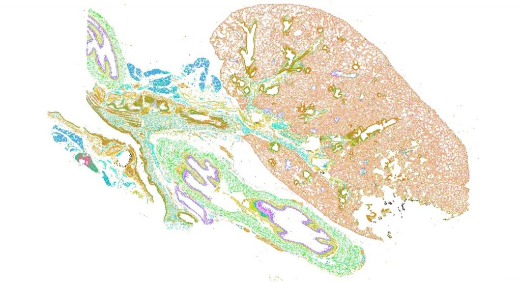
This cross-section of a mouse lung was analyzed using spatial transcriptomics. The cells have been colored by type.
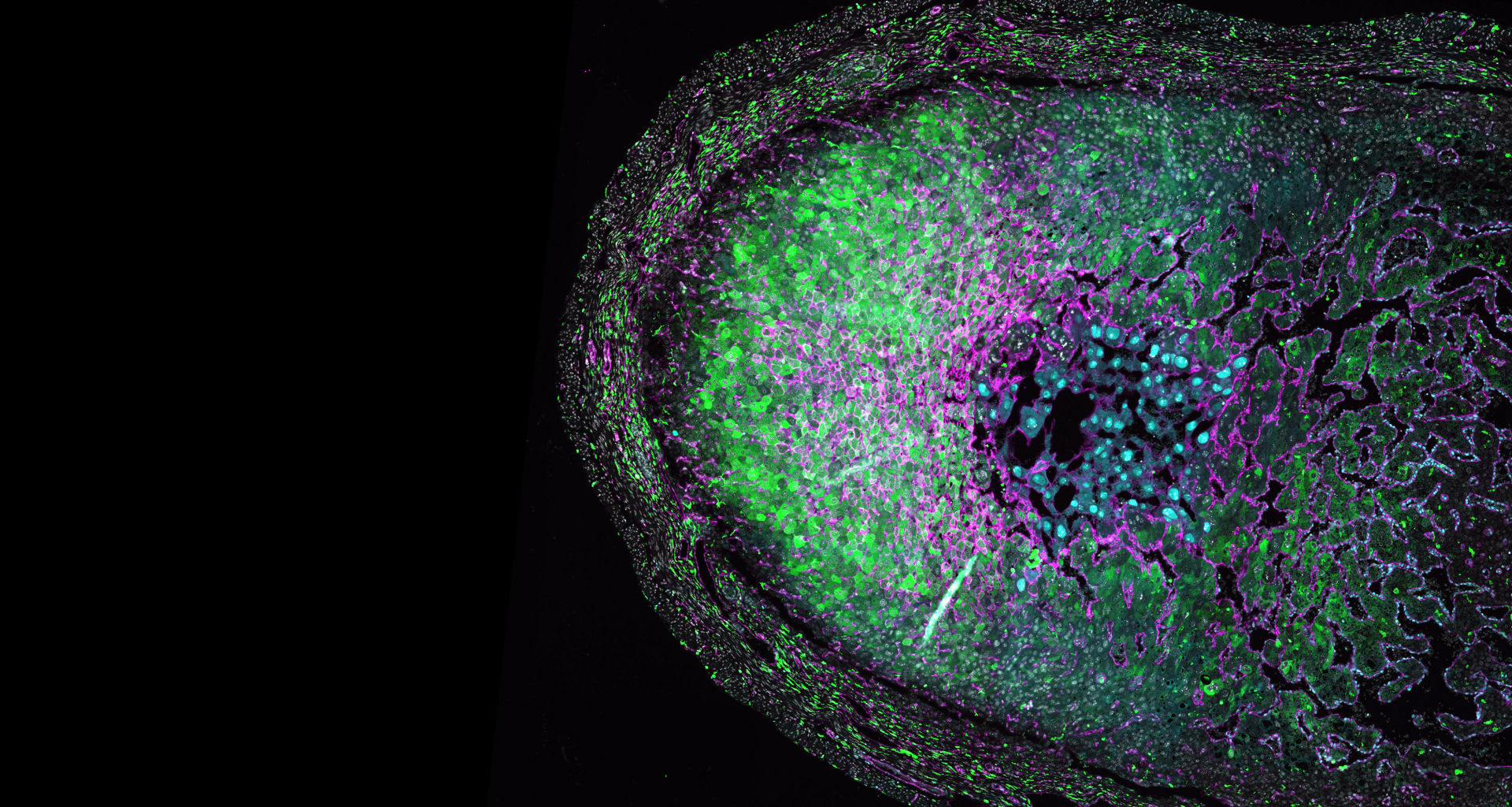
The maternal-fetal interface
Few tissue environments are as complex as the maternal-fetal interface, the site at which a parent’s immune cells interact with rapidly developing tissues—and immune cells—belonging to a fetus.
In this cross-section of a mouse embryo at day 8.5 of gestation, the cells have been stained to display markers associated with immune cells (green) and epithelial cells (pink). This image helps shed light on the basic biology of pregnancy and gives researchers clues to how immune cells defend a developing fetus.
These stunning images were captured at LJI using cutting-edge Orion microscopy equipment and software, funded by LJI donors Michael and Ellise Coit.